M3D Cell Culture Kits Greiner Bio-One


Magnetic M3D Cell Culture
The core technology of Greiner Bio-One’s Magnetic M3D Cell Culture is the magnetisation of cells with NanoShuttle™-PL. The cells can be aggregated with magnetic forces, either by levitation or printing, to form structurally and biologically representative M3D models in vitro. NanoShuttle™-PL consists of gold, iron oxide, and Poly-L-Lysine. These nanoparticles (Ø < 50 nm) magnetise cells by electrostatically attaching
to cell membranes during an overnight static incubation. Magnetised cells will appear peppered with dark nanoparticles after incubation. NanoShuttle™-PL is biocompatible, having no effect on metabolism, proliferation and inflammatory stress. Additionally, it does not interfere with experimental techniques,
such as fluorescence or Western blotting. With magnetised spheroids, solution addition and removal is made easy by using magnetic force to hold them in a stationary position during aspiration, thereby limiting spheroid loss. Spheroids can also be picked up and transferred between vessels using magnetic tools such as the MagPen™ (7.657 850).
Advantages of magnetic cell culture:
- M3D in a 2D workflow
- Fast M3D tissue assembly
- Easy to handle / no sample loss
- Scalable – single well to 384 well
- Ease of co-culturing cells
- High-throughput Screening (HTS) with flat bottom for optical imaging
- Ready for automation
- Positioning spheroid centrally within the well
Magnetic Levitation
Magnetic levitation is an easy tool to create native tissue environments in vitro. Cells are magnetised with
NanoShuttle™-PL through overnight incubation and dispensed into a cell-repellent dish or multiwell plate, where they are levitated off the bottom by a magnet above the cell-repellent vessel. When levitating, cells come off the stiff well bottom and the magnetic forces work as an invisible scaffold that rapidly aggregates cells to encourage cell-cell interactions and induce ECM synthesis. The magnetic levitation method has been successfully used to make M3D cultures with different cell types, including cell lines, stem cells and primary cells. The basic application of this technology is to culture M3D cell cultures under different environmental conditions and then analyse them using common biological research techniques, such as
immunohistochemical analysis and western blotting.
Magnetic Bioprinting
In contrast to magnetic levitation, with magnetic 3D bioprinting, cells incubated with NanoShuttle™-PL overnight are printed into spheroids by placing a microplate containing magnetised cells atop a drive of magnets. The magnets below each well utilise mild magnetic forces to induce cell aggregation and print a spheroid at the bottom of each well. After 15 minutes to a few hours, the plate containing the spheroids can be removed from the magnetic drive and cultured long-term without use of magnetic force. This
system overcomes the limitations of other platforms by enabling rapid formation of spheroids unattached to any stiff substrate, reproducible in size without limitation to cell type and scalable in size for high-throughput formats (96 and 384 well). With magnetic 3D bioprinting, viable spheroids can be printed that compact and subsequently grow over time, facilitating continuous assessment of cell viability and other functions using commercially available assays. The 3D printing method, together with commercially
available standardised assay methods, provide an ideal combination for high-throughput compound screening.
Magnetic Printing of Rings and Imaging System
Additionally, it was demonstrated that magnetic 3D bioprinting can be used to develop novel migration assays (ring formation) and that automated kinetic imaging can facilitate high-throughput screening. These assays form a foundation for robust screening of compound effects on cell migration. Based on magnetic 3D bioprinting, cells magnetised with NanoShuttle™-PL are printed into 3D rings. Immediately after printing, these structures will compact and eventually close, as a function of cell migration and viability. Ring closure can be captured using a compact imaging kit with an iPod*) programmed by a freely available app (Experiment Assistant) to image whole plates at specific intervals, foregoing the need
to image well-by-well under a microscope (Fig. 4). Culture contraction is generally complete within 24 hours, and images are batch processed to rapidly yield toxicity data. Moreover, as closure is label-free, the remaining rings are available for further experimentation (IHC, Western blot, genomics, etc.).
| 7.655 830 | 96-Well Drives |
• 96-Well Spheroid Drive (1) • 96-Well Holding Drive (1) |
CHF 1'727.55
|
|
||
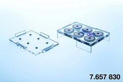
| 7.657 830 | 6-Well Drives |
• 6-Well Levitating Drive (1) • 6-Well Concentrating Drive (1) |
CHF 1'485.10
|
|
||
| 7.662 830 | 24-Well Drives |
• 24-Well Levitating Drive (1) • 24-Well Concentrating Drive (1) |
CHF 1'636.68
|
|
||
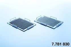
| 7.781 830 | 384-Well Drives |
• 384-Well Spheroid Drive (1) • 384-Well Holding Drive (1) |
CHF 2'549.08
|
|
Maybe you also like...


Recent products
Follow us
Our Newsletter
Subscribe now